Science Anatomy and Physiology Anatomy and Physiology questions and answers Correctly label the following anatomical features of a nerve. Anterior root Spinal nerve Posterior root Posterior root Blood vessels Reset Zoom < Prev 20 of 50 Ne, > F6 8% 2 3 4 5 9 This problem has been solved!
PHYSICAL ASSESSMENT- EYE & EAR
Correctly label the following anatomical features of a neuron. … Correctly label the following anatomical features of a nerve. Each of the labels below describes a sensory or motor innervation. Identify the spinal nerve plexuses pictured below and drag the innervations to the appropriate category according to which plexus is responsible.

Source Image: coursehero.com
Download Image
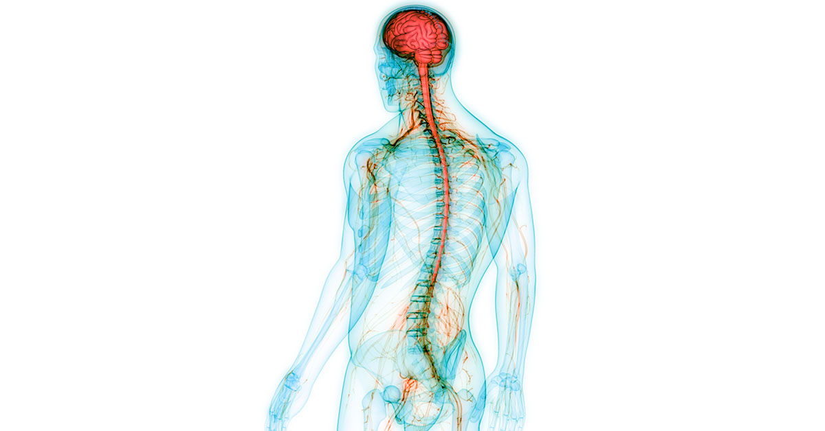
Like the heart, lungs, and stomach, the nervous system is made up of specialized cells. These include nerve cells (or neurons) and glial cells (or glia ). Neurons are the basic functional units of the nervous system, and they generate electrical signals called action potentials, which allow them to quickly transmit information over long
Source Image: wholehealthsource.blogspot.com
Download Image
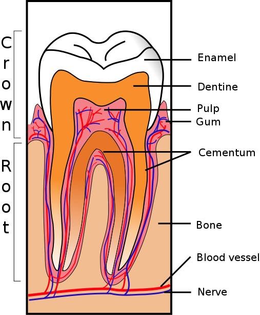
Stream [PDF] Download Medical Terminology & Anatomy for Coding Free download and Read by Luvina Trust | Listen online for free on SoundCloud Nov 28, 2022The perineurium lies within the nerve and consists of a protective sheath that surrounds bundles of nerve fibers, termed fascicles. On the other hand, nerve roots connect the central nervous system to the peripheral nerves. The posterior and anterior roots are components of each spinal nerve, with the posterior root housing a posterior root

Source Image: coursehero.com
Download Image
Correctly Label The Following Anatomical Features Of A Nerve.
Nov 28, 2022The perineurium lies within the nerve and consists of a protective sheath that surrounds bundles of nerve fibers, termed fascicles. On the other hand, nerve roots connect the central nervous system to the peripheral nerves. The posterior and anterior roots are components of each spinal nerve, with the posterior root housing a posterior root Correctly label the following anatomical features of the cerebellum. Correctly label the following functional regions of the cerebral cortex. Consider a situation where a stroke or mechanical trauma has occurred resulting in damage to one of the areas of the brain indicated in the image.
1FA31D70-247B-4691-99E1-7C24A8844E69.jpeg – Correctly label the following anatomical features of a neuron | Course Hero
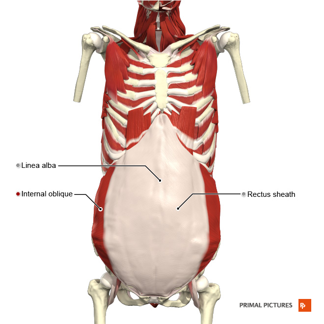
Correctly label the following anatomical features of a nerve. Anterior root R ootlets Endoneurium Endoneurium Blood vessels Blood vessels Unmyelinated nerve fibers Unmyelinated nerve Myelin Epineurium Spinal nerve Posterior root ganglion Fascicle (8 01:57:44 Perineurium This problem has been solved! Muscles of Respiration – Physiopedia
Source Image: physio-pedia.com
Download Image
Understanding the Higher-Order Approach to Consciousness: Trends in Cognitive Sciences Correctly label the following anatomical features of a nerve. Anterior root R ootlets Endoneurium Endoneurium Blood vessels Blood vessels Unmyelinated nerve fibers Unmyelinated nerve Myelin Epineurium Spinal nerve Posterior root ganglion Fascicle (8 01:57:44 Perineurium This problem has been solved!

Source Image: cell.com
Download Image
PHYSICAL ASSESSMENT- EYE & EAR Science Anatomy and Physiology Anatomy and Physiology questions and answers Correctly label the following anatomical features of a nerve. Anterior root Spinal nerve Posterior root Posterior root Blood vessels Reset Zoom < Prev 20 of 50 Ne, > F6 8% 2 3 4 5 9 This problem has been solved!

Source Image: thenurseslab.com
Download Image
Stream [PDF] Download Medical Terminology & Anatomy for Coding Free download and Read by Luvina Trust | Listen online for free on SoundCloud Like the heart, lungs, and stomach, the nervous system is made up of specialized cells. These include nerve cells (or neurons) and glial cells (or glia ). Neurons are the basic functional units of the nervous system, and they generate electrical signals called action potentials, which allow them to quickly transmit information over long
![Stream [PDF] Download Medical Terminology & Anatomy for Coding Free download and Read by Luvina Trust | Listen online for free on SoundCloud](https://i1.sndcdn.com/artworks-ffEjFv6MV8TVV7LL-B3E0wA-t500x500.jpg)
Source Image: soundcloud.com
Download Image
What Do the Different Parts of The Nervous System Do? – Regional Neurological Associates Obtain slides of each of the following tissues, observe them, draw and label the significant features. SLIDES: Spinal cord, cerebral cortex, cerebellum, dorsal root ganglion, peripheral nerve, neuromuscular junction: 1. Obtain a slide of nervous tissue from the slide box.
Source Image: regionalneurological.com
Download Image
Think Tank Centre Nov 28, 2022The perineurium lies within the nerve and consists of a protective sheath that surrounds bundles of nerve fibers, termed fascicles. On the other hand, nerve roots connect the central nervous system to the peripheral nerves. The posterior and anterior roots are components of each spinal nerve, with the posterior root housing a posterior root

Source Image: thinktankcentre.blogspot.com
Download Image
Anatomy of the Urinary System Correctly label the following anatomical features of the cerebellum. Correctly label the following functional regions of the cerebral cortex. Consider a situation where a stroke or mechanical trauma has occurred resulting in damage to one of the areas of the brain indicated in the image.
Source Image: stanfordchildrens.org
Download Image
Understanding the Higher-Order Approach to Consciousness: Trends in Cognitive Sciences
Anatomy of the Urinary System Correctly label the following anatomical features of a neuron. … Correctly label the following anatomical features of a nerve. Each of the labels below describes a sensory or motor innervation. Identify the spinal nerve plexuses pictured below and drag the innervations to the appropriate category according to which plexus is responsible.
Stream [PDF] Download Medical Terminology & Anatomy for Coding Free download and Read by Luvina Trust | Listen online for free on SoundCloud Think Tank Centre Obtain slides of each of the following tissues, observe them, draw and label the significant features. SLIDES: Spinal cord, cerebral cortex, cerebellum, dorsal root ganglion, peripheral nerve, neuromuscular junction: 1. Obtain a slide of nervous tissue from the slide box.
